
Honest, Unambiguous
and
Updated
technical information for Stevia Industries

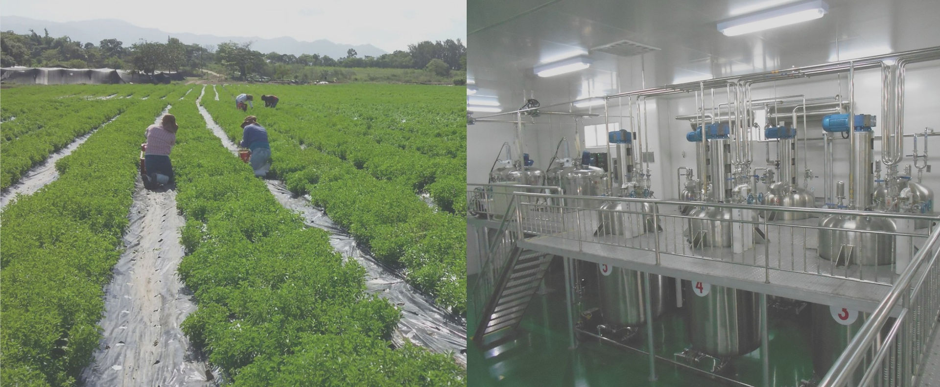
Farming

Extraction

Formulation


Stevia Tissue Culture Using Leaves As Explants
Often, portions of leaf lamina are used as explants for tissue culture of Stevia. Explants are generally excised from young leaf primordia or immature young leaf of the shoot apex. Depending on the explant size and its nature, and culture conditions, the explant may grow into an undifferentiated callus or may directly grow adventitious shoots and roots. Stevia leaf explants, when grown in presence of high kinetin concentration, are reported to grow shoot buds at the margins of the explants. Higher concentration of Auxins favour callus formation from the explants. Excised portions of the callus, when transferred to media containing higher amount of cytokinins, grow adventitious shots. Rhizogenesis in those shoots can be triggered by transfer into media containing appropriate levels of auxins.
The following table shows some of the work done on Stevia tissue culture using leaves as explant source.


Regeneration of multiple shoots from leaf derived callus of Stevia rebaudiana. (A) Callus initiation of leaf explant. (B & C) Proliferation of multiple shoots from leaf derived callus at day 28.
From:
Janarthanam, B. & Gopalakrishnan, M. & Sai, G. & Sekar, T.. (2009). Plant Regeneration from Leaf Derived Callus of Stevia rebaudiana Bertoni. Plant Tissue Culture and Biotechnology. 19(2): 133-141
De novo shoot regeneration from leaf explant of Stevia rebaudiana in MS medium supplemented with 30 g dm-3 sucrose, 8.88 μM BA and 4.65 μM Kn. A - Knob like shoot buds on either side of midrib 4 weeks after inoculation (M - midrib, bar = 2.5 mm). B - Further growth and greening of shoot bud 5 weeks after inoculation (bar = 2.5 mm). C - A pair of green leaves emerging from shoot bud (bar = 2.5 mm). D - Transverse section of leaf showing multiple shoot on leaf surface 4 weeks after inoculation (bar = 0.11 mm). E - Transverse section of leaf showing a group of cells organizing to form shoot primordia (V - vasculature, bar = 0.11 mm). F - Transverse section of leaf showing more organized shoot bud with its own vasculature 5 weeks after inoculation (V - vasculature, bar = 0.11 mm).
From:
Sreedhar RV, Venkatachalam L, Thimmaraju R, Bhagyalakshmi N, Narayan MS and G.A. Ravishankar GA (2008) Direct organogenesis from leaf explants of Stevia rebaudiana and cultivation in bioreactor; Biologia Plantarum 52 (2): 355-360, 2008

Stage of micropropagation of Stevia rebaudiana Bert. (a) Leaf Explants showing callus initiation response; (b) Explant derived Callus showing growth and shoot differentiation; (c) Differentiated shoots are growing for shoot elongation
From:
Roy Pathak, Malabika. (2016). An Efficient Organogenesis Based Micropropagation of Medicinal and Natural Sweet Leave Plant Stevia Rebaudiana Bert. in Kingdom of Bahrain. IOSR Journal of Biotechnology and Biochemistry (IOSR-JBB). 2. 91-97.